Top Doctors editorial
Created by: Top Doctors editorial
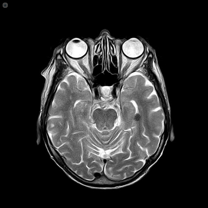
A brain MRI is a type of scan which uses a magnetic field and radio waves to produce detailed images of your brain. An MRI is carried out by a radiologist, and is a safe and pain-free procedure.
Why do I need a brain MRI?
A brain MRI is used to investigate a number of things. If you have gone to the doctor with symptoms such as dizziness, headaches, seizures, or changes in your behaviour, a brain MRI can help to check if there are problems with the tissues in your brain. A brain MRI is also often used to determine if there is any damage to the brain after an injury or stoke, and if so, what kind of damage.
The images produced from the scan can help to diagnose the following conditions:
- aneurysms
- stroke
- haemorrhage
- multiple sclerosis
- tumours
- spinal cord injuries
- infections
- brain trauma
If the MRI machine is used to examine the blood vessels, the test is instead known as a Magnetic Resonance Angiography (MRA). However, the test still involves exactly the same process.
You might need to have further scans or tests after the brain MRI before a formal diagnosis can be given.
Functional MRI scans
A functional MRI of the brain (fMRI) is a special kind of brain MRI used to diagnose dementia, assess damage from a head injury, or prepare for brain surgery. The fMRI involves scanning different parts of the brain to determine which part performs which functions, in particular important communication and movement functions. To determine this you will be asked to perform basic tasks, such as answering questions, and the scan will detect what is happening in your brain while you carry these tasks out.
What to expect in a brain MRI
For a detailed look at how MRI tests work, what you can expect, and how to prepare, see our page on MRI scans.
In a brain MRI, the test generally takes between 30 and 60 minutes. You might have a plastic coil placed around your head, and you might be administered a special called gadolinium through an IV. This can help doctors understand the images from the scan by highlighting different areas of the brain, particularly the blood vessels. The dye is usually safe but there may be a risk if you have problems with your kidney. You should make your doctor aware of any pre-existing conditions before they give you the IV.